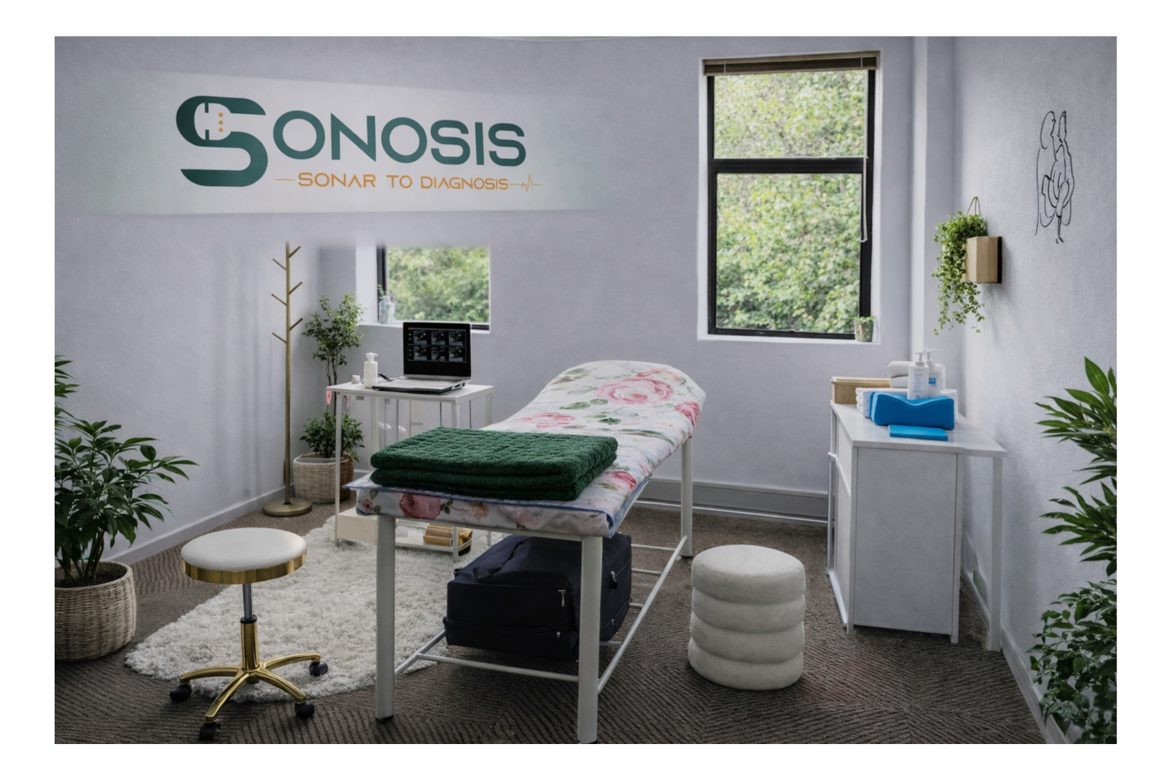
Diagnostic Ultrasound You Can Trust
Accurate imaging. Compassionate care. Same-day reporting.
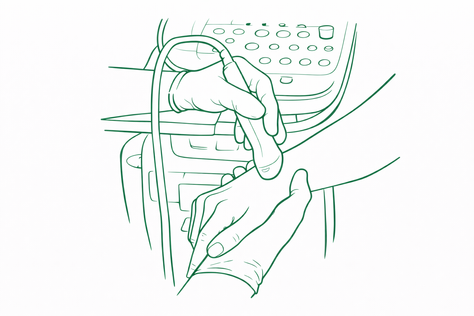
Performed by a qualified diagnostic sonographer using advanced ultrasound technology — without long waiting times.
Why choose Sonosis Ultrasound?
At Sonosis, we believe diagnostic imaging should be accurate, accessible, and patient-centred.
We offer high-quality ultrasound examinations performed by an experienced diagnostic sonographer, with a strong focus on clinical detail, clear reporting, and compassionate care.
What sets Sonosis apart:
• Same-day scanning and prompt reporting
• Personal, one-on-one care — no rushed appointments
• High-resolution diagnostic ultrasound
• In-practice and mobile ultrasound services
• Reduced waiting times compared to hospital or radiology practices
• Affordable pricing with transparent fees
• Close collaboration with referring doctors and allied health professionals
Whether you are coming in for a routine scan, a pregnancy ultrasound, or a focused musculoskeletal assessment, our goal is simple — to provide accurate answers and peace of mind.
Sonosis — from sonar to diagnosis.
Our ultrasound services include:
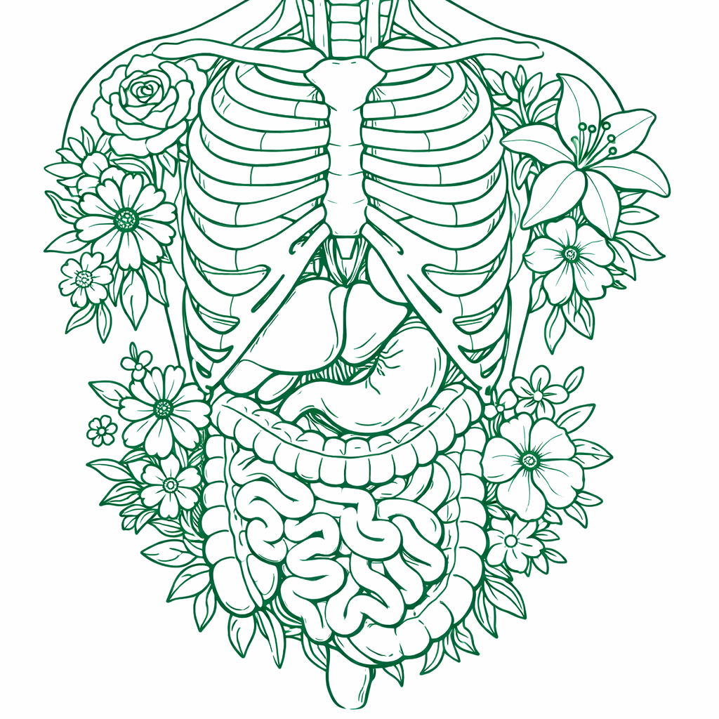
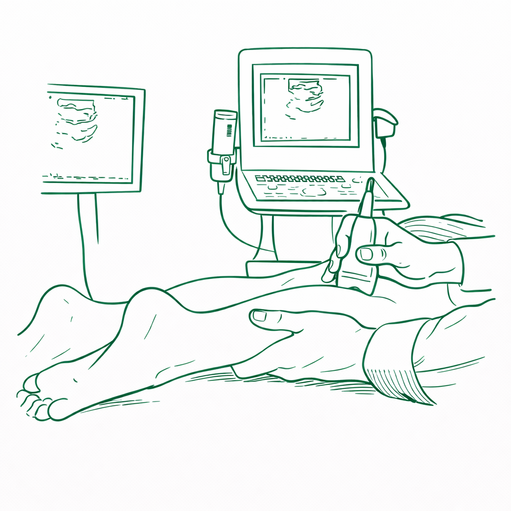
An abdomen and pelvic ultrasound is a painless, non-invasive examination that uses sound waves to assess the organs inside the abdomen and pelvis.
This scan may evaluate the following:
Abdominal organs:
Liver
Gallbladder and bile ducts
Pancreas
Spleen
Kidneys
Major abdominal blood vessels
Pelvic organs (female):
Uterus
Endometrium
Ovaries
Adnexal regions
Pelvic organs (male):
Bladder
Prostate (transabdominal view)
Preparation instructions
To ensure the best image quality, correct preparation is very important.
Fasting (for abdominal assessment):
Do not eat for 6–8 hours before the scan
You may drink small amounts of plain water only
No tea, coffee, milk, or fizzy drinks
Full bladder (for pelvic assessment):
Drink 1–1.5 litres of water about 1 hour before the appointment
Do not empty your bladder until after the scan
A full bladder helps improve visualisation of the pelvic organs.
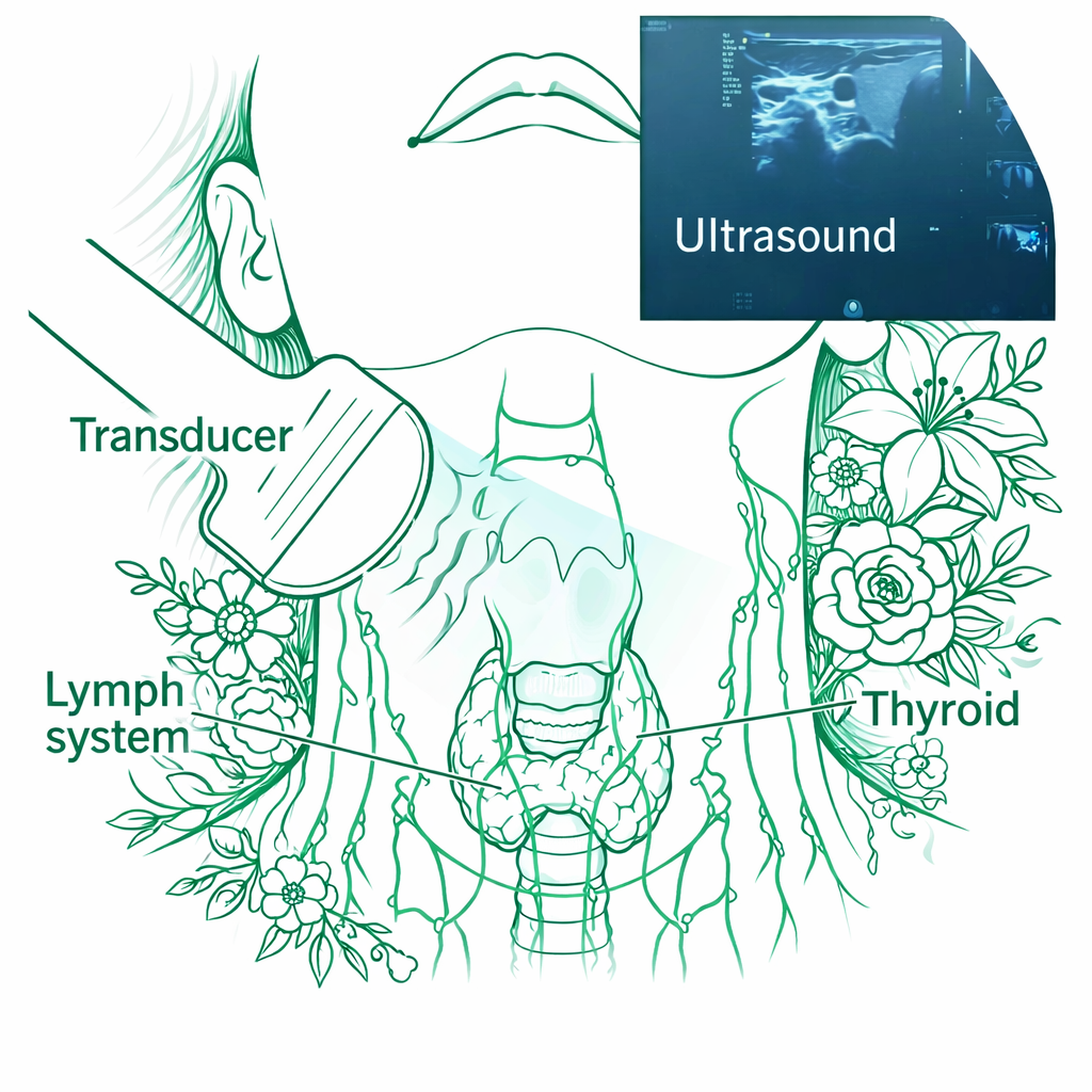
Thyroid & Neck Ultrasound
A thyroid and neck ultrasound is a diagnostic imaging examination used to assess the thyroid gland and surrounding neck structures.
The examination is performed by a registered diagnostic sonographer and provides a formal diagnostic report of findings for your referring doctor or healthcare provider.
Structures assessed include:
Thyroid gland (size, texture, and nodules)
Parathyroid region (when indicated)
Cervical lymph nodes
Surrounding soft tissues of the neck
Thyroid nodules are carefully evaluated and, where appropriate, classified using standard TI-RADS criteria to assist your doctor in clinical decision-making.
Common reasons for referral include:
Thyroid nodules or swelling
Enlarged thyroid (goitre)
Abnormal thyroid blood results
Neck lumps
Follow-up of known thyroid conditions
Preparation instructions
No fasting required
No special preparation needed
You may eat, drink, and take medication as normal
Important note
This is a diagnostic medical examination. A written ultrasound report is provided following the scan and may be used by your doctor to guide further investigation, follow-up, or treatment.
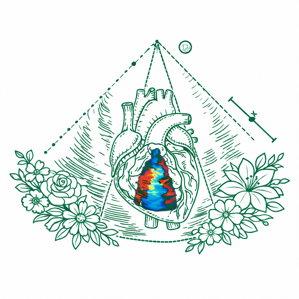
Vascular & Doppler Ultrasound Studies
Vascular and Doppler ultrasound studies are diagnostic examinations used to assess blood flow within the heart, arteries, and veins.
These studies are performed by a registered diagnostic sonographer and provide a formal diagnostic report of findings for your referring doctor or healthcare provider.
Doppler ultrasound evaluates blood flow direction, velocity, and waveform patterns, assisting in the diagnosis of circulatory and cardiovascular conditions.
Types of Doppler studies performed include:
Cardiac (Heart) Ultrasound
Assessment of heart chambers and valves
Evaluation of cardiac function
Doppler assessment of blood flow through the heart
Arterial Doppler Studies
Upper or lower limb arterial circulation
Assessment of arterial narrowing or blockages
Investigation of poor circulation, pain, or claudication
Venous Doppler Studies
Evaluation for deep vein thrombosis (DVT)
Assessment of venous patency and flow
Investigation of limb swelling or pain
Venous Insufficiency & Varicose Vein Studies
Mapping of superficial and deep venous systems
Assessment of venous reflux
Evaluation of chronic venous insufficiency and varicose veins
Common indications include:
Suspected blood clots (DVT)
Leg or arm swelling
Pain related to circulation problems
Varicose veins
Stroke risk screening (carotid arteries)
Known cardiovascular or peripheral vascular disease
Preparation instructions
No fasting required unless specifically advised
Wear loose, comfortable clothing
Shorts may be helpful for lower limb studies
Important note
All vascular and Doppler studies performed at Sonosis Ultrasound are diagnostic medical examinations. A detailed ultrasound report is provided to assist your doctor in further management, referral, or treatment planning.
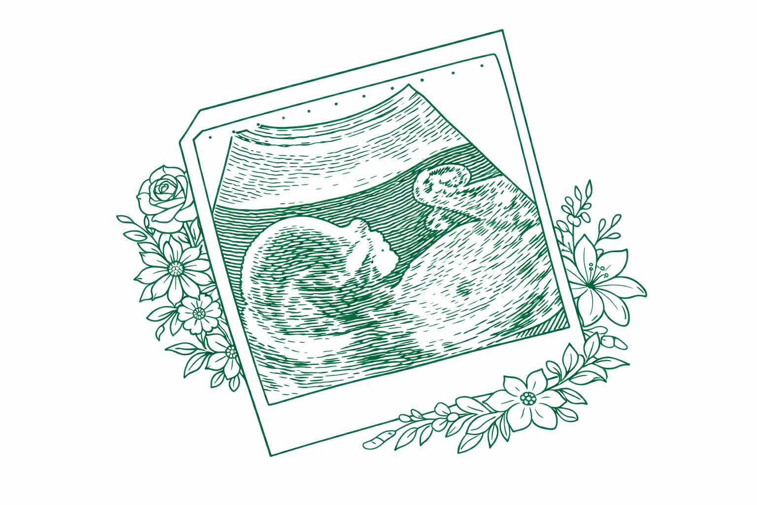
Pregnancy ultrasound is a safe, non-invasive imaging examination used to assess the developing pregnancy and monitor your baby’s growth and wellbeing.
At Sonosis, we understand how important reassurance and clarity are during pregnancy. Our scans are performed with care, accuracy, and respect for both mother and baby.
Depending on the stage of pregnancy, ultrasound can help to:
• Confirm an intra-uterine pregnancy
• Estimate gestational age and expected due date
• Assess fetal heartbeat and early development
• Evaluate fetal growth and anatomy
• Assess placental position and amniotic fluid
• Investigate pain, bleeding, or other pregnancy-related concerns
All scans are performed using high-resolution ultrasound equipment, with same-day reporting and clear explanations provided during your appointment.
Please note that pregnancy ultrasounds are diagnostic examinations. While we aim to make the experience as comfortable and reassuring as possible, these scans are focused on medical assessment rather than entertainment imaging.
If you have been referred by your doctor or require reassurance during pregnancy, we are here to support you every step of the way — from sonar to diagnosis.
Musculoskeletal & Sports Injury Ultrasound
A musculoskeletal (MSK) ultrasound is a diagnostic imaging examination used to assess muscles, tendons, ligaments, joints, bursae, and surrounding soft tissues.
It is particularly useful for evaluating sports injuries, acute trauma, overuse injuries, and chronic pain, allowing real-time assessment of both structure and movement.
This examination is performed by a registered diagnostic sonographer and provides a detailed diagnostic report of findings for your referring doctor or healthcare provider.
Common conditions assessed include:
Tendon tears (partial or full thickness)
Muscle strains and ruptures
Ligament injuries
Rotator cuff pathology
Tennis or golfer’s elbow
Achilles and patellar tendon injuries
Bursitis
Joint effusions
Soft tissue masses or swelling
Dynamic assessment may be performed, allowing evaluation of structures during movement, which is especially helpful in sports-related injuries.
Advantages of musculoskeletal ultrasound
No radiation
Real-time imaging
Dynamic assessment during movement
Ability to compare with the opposite side
Excellent evaluation of superficial soft tissues
Preparation instructions
No fasting required
Please wear comfortable clothing or bring shorts / vest if the shoulder, hip, knee, or ankle is being assessed
Important note
This is a diagnostic medical examination. A written ultrasound report is provided following the scan and may assist your doctor or therapist in guiding further management or treatment.
Soft tissue ultrasound is a safe, non-invasive imaging examination used to evaluate lumps, swellings, and areas of localised pain beneath the skin.
At Sonosis, we frequently assess soft tissue concerns such as palpable lumps, swelling, tenderness, or changes noticed by the patient or referring clinician. Ultrasound allows real-time assessment of the underlying structures without radiation.
Soft tissue ultrasound can help to assess:
• Lumps and swellings beneath the skin
• Lipomas (fatty lesions)
• Cysts and fluid collections
• Inflamed or infected tissue
• Enlarged or reactive lymph nodes
• Post-traumatic swelling or haematomas
• Localised pain of unknown origin
Using high-resolution ultrasound, we are able to differentiate between cystic and solid structures, assess vascularity when needed, and guide further management if required.
All examinations are performed with care and discretion, with same-day reporting and clear communication of findings.
If a lump or area of concern has changed in size, become painful, or is causing anxiety, ultrasound is often the first and most appropriate imaging step.
From assessment to reassurance — Sonosis is here to help guide you toward clarity and diagnosis.
Services we offer
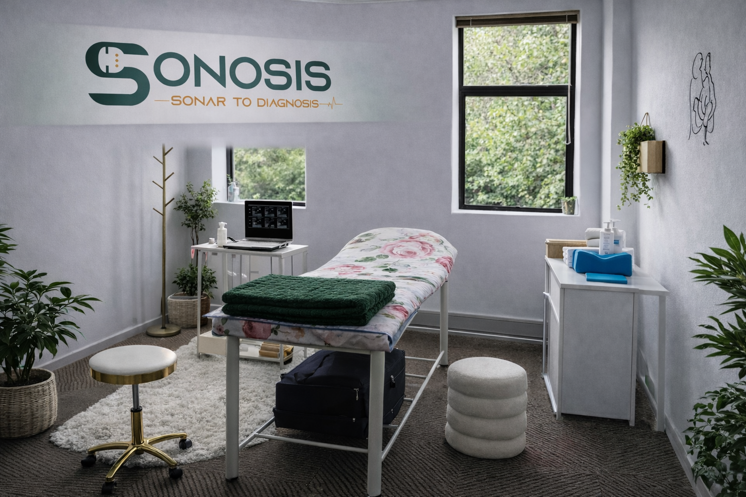
We offer in-house diagnostic ultrasound scanning at the following Sonosis branches, allowing for convenient access, prompt assessment, and same-day reporting.

Our mobile ultrasound service is ideal for patients who are unable to visit one of our branches, or who prefer the comfort and privacy of their own home.
We bring professional, high-quality diagnostic ultrasound imaging directly to you.
A mobile service fee applies and is calculated based on distance from the nearest Sonosis branch at AA travel rates, with an additional R500 added to the standard scan fee.
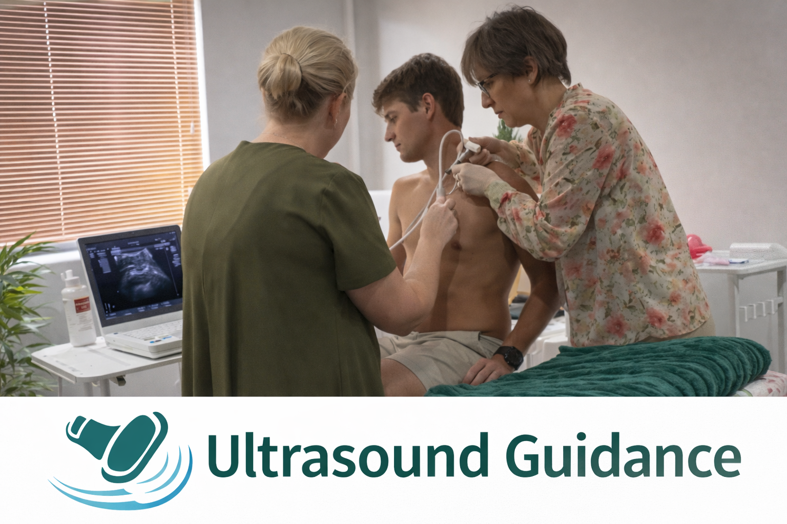
Sonosis provides high-precision ultrasound guidance for interventional and diagnostic procedures, ensuring accurate needle placement and real-time visualisation of target structures.
Using high-resolution imaging and dynamic guidance, procedures are performed with enhanced safety, improved anatomical confidence, and reduced risk to adjacent nerves, vessels, and soft tissues.
Ultrasound guidance allows for:
Precise targeting of joints, tendons, bursae, ligaments, nerves, and peri-articular structures
Continuous real-time needle visualisation
Optimised delivery of therapeutic agents to the intended anatomical plane
Improved procedural accuracy and patient comfort
Our service supports a wide range of musculoskeletal, pain-management, and regenerative procedures, including:
Joint and peri-articular injections
Bursal and tendon sheath injections
Ligament and enthesis targeting
Nerve hydrodissection and peri-neural guidance
Complex multi-level and layered injection planning
All guidance is performed by an experienced diagnostic sonographer with advanced anatomical expertise, ensuring high-precision, reproducible, and clinically reliable procedural support.
Follow Us
⦁
Follow Us
Follow Us ⦁ Follow Us


Patient Reviews
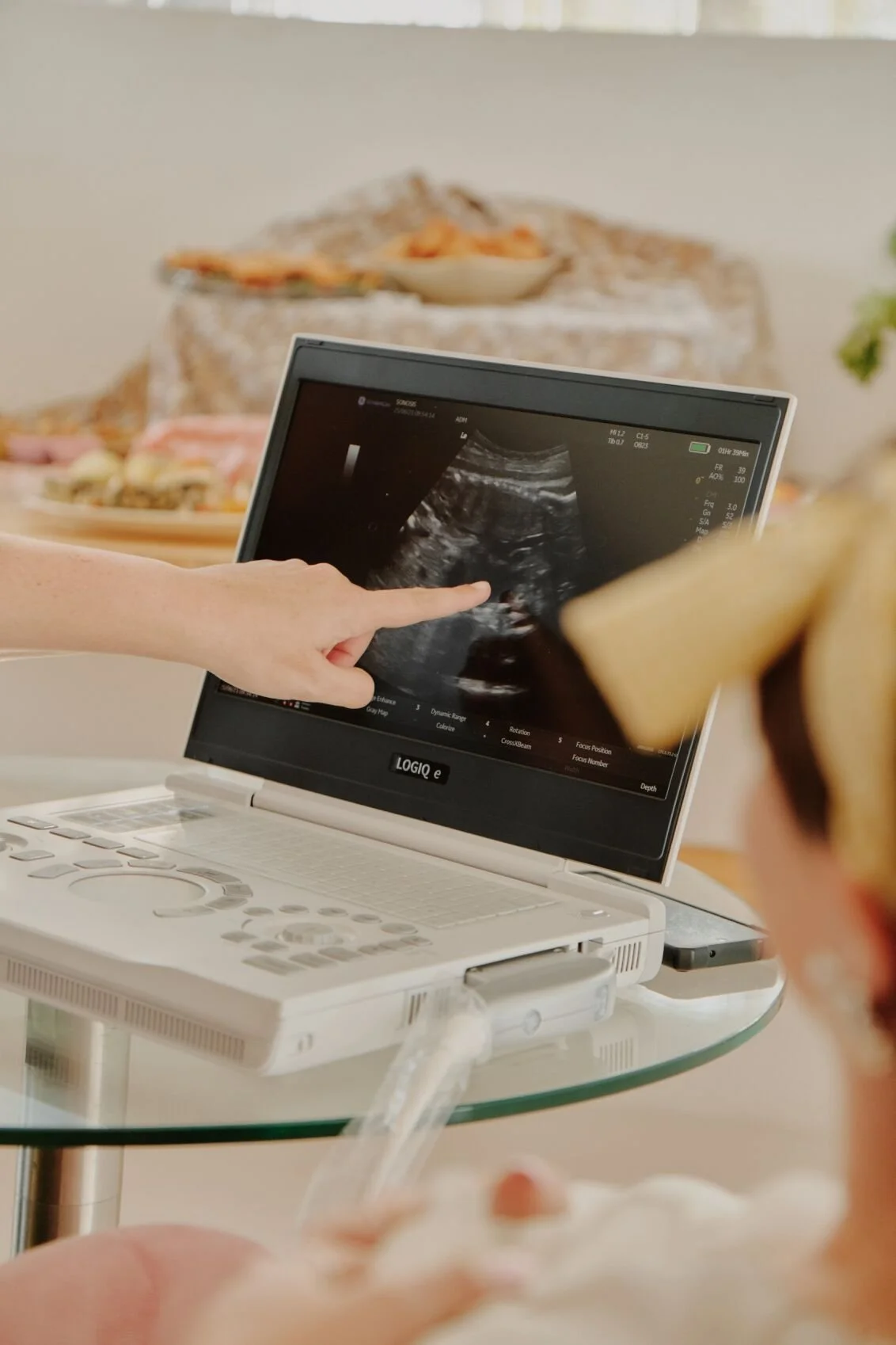
We were so blessed to have Caroline come to our baby shower and make the day even more special! She was so kind, gentle, and professional, and she made everyone feel included and excited. Seeing our little one on the screen surrounded by family and friends was such a magical moment, something we’ll never forget. She explained everything so clearly and made the whole experience full of joy and love.
Thank you for helping us create such beautiful memories. We can’t recommend her enough!
— Anelda Hartung Visagie“I’m so happy with Caroline Van Graan’s service after struggling to check for my baby’s gender it took just one visit to her practice and she was able to pick up the gender of my baby. Her service was excellent not to mention taking us through every step of what she was doing, and she’s so patient and passionate when doing her job.”
— Zintle Mlenze“Very convenient and efficient to be able to conduct ultrasound exam at medical rooms in Randburg instead of needing to go to a hospital and fill out forms. Report was prompt and thorough. Very happy with Caroline’s service.”
— Greg Brady Contact Us
Eager to explore a collaboration? Kindly share your details, and we will reach out to you promptly. We look forward to the opportunity to connect.